AB-Abdomen Exam Questions & Answers
Abdomen Sonography Examination • ARDMS
100% money-back guarantee
Sample AB-Abdomen Questions
Practice with real exam-style questions, each with the verified correct answer and explanation.
Which technique is best for demonstrating the characteristic of the small hepatic lesion identified by the arrow on this image?
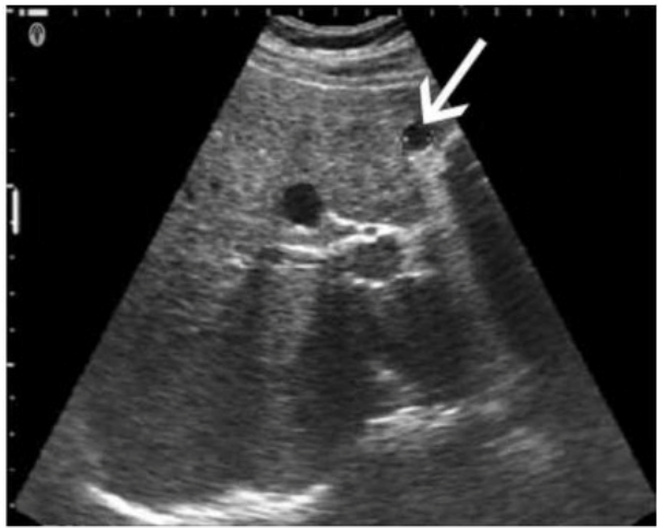
The image shows a small hepatic lesion located very close to the anterior liver capsule, as indicated by the arrow. When imaging very superficial or near-field structures like subcapsular hepatic lesions, using a standoff pad is the most effective technique for optimizing visualization.
A standoff pad (also known as an acoustic stand-off or gel pad) helps increase the distance between the transducer and the superficial target. This improves the focus and beam shape for near-field imaging and minimizes reverberation and ring-down artifacts. It allows better evaluation of superficial lesions by positioning them within the focal zone of the transducer, which is usually set a few millimeters below the probe surface.
Differentiation from other options:
A . Decrease depth: While reducing depth can help center deeper lesions in the field of view, it does not address issues with near-field resolution.
B . Scan in upright position: This may help in gallbladder or fluid positioning but is not optimal for improving visualization of superficial liver lesions.
C . Move the transducer focus: Adjusting focus deeper into the image won't enhance resolution of very superficial structures unless a standoff is used to bring the lesion into the focal zone.
Rumack CM, Wilson SR, Charboneau JW, Levine D. Diagnostic Ultrasound. 5th Edition. Elsevier, 2018. Chapter: Liver, pp. 80--84.
Kremkau FW. Sonography: Principles and Instruments. 9th Edition. Elsevier, 2015. Chapter: Image Formation and Optimization, pp. 114--117.
AIUM Practice Parameter for the Performance of an Ultrasound Examination of the Abdomen and/or Retroperitoneum, 2020.
Which structure is most likely shown in this image of the right lower quadrant?
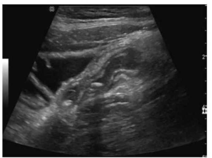
The ultrasound image shows a blind-ending, non-compressible, tubular structure in the right lower quadrant with a target or bullseye appearance in transverse section --- highly suggestive of the appendix.
Sonographic features of the appendix (especially in suspected appendicitis):
Blind-ending tubular structure arising from the cecum
Non-compressible on graded compression
Diameter >6 mm is suggestive of appendicitis
May demonstrate a ''target sign'' in transverse view (concentric ring-like appearance)
Increased echogenicity of surrounding fat in cases of inflammation
May contain an appendicolith or show hyperemia on color Doppler if inflamed
The location (right lower quadrant) and appearance in this case are classic for the normal or potentially inflamed appendix.
Differentiation from other options:
A . Fallopian tube: Located more in the adnexal regions and usually not visible unless distended (e.g., hydrosalpinx).
B . Ureter: Usually not visualized on ultrasound unless dilated due to obstruction.
D . Jejunum: Has valvulae conniventes ('keyboard sign') and peristalsis; does not present with a blind-ending tubular appearance from the cecum.
Rumack CM, Wilson SR, Charboneau JW, Levine D. Diagnostic Ultrasound. 5th Edition. Elsevier, 2018. Chapter: Gastrointestinal Tract, pp. 460--468.
American College of Radiology (ACR). ACR Appropriateness Criteria --- Right Lower Quadrant Pain -- Suspected Appendicitis.
AIUM Practice Parameter for the Performance of a Pediatric Abdominal and/or Retroperitoneal Ultrasound Examination, 2020.
A patient presents with ampulla of Vater obstruction, distention of the gallbladder, and painless jaundice. Which condition is most likely associated with these findings?
Courvoisier sign describes the clinical finding of painless jaundice combined with a palpable, distended gallbladder. This typically results from obstruction at the distal common bile duct, often due to pancreatic head carcinoma or cholangiocarcinoma, leading to bile accumulation and gallbladder distention. In contrast, Mirizzi syndrome involves compression of the common hepatic duct by an impacted stone in the cystic duct.
According to Rumack's Diagnostic Ultrasound and standard clinical references:
''Courvoisier sign refers to a palpable, enlarged gallbladder due to obstruction of the distal bile duct, often from malignancy.''
Rumack CM, Wilson SR, Charboneau JW, Levine D. Diagnostic Ultrasound. 5th ed. Elsevier, 2017.
Moore KL. Clinically Oriented Anatomy. 8th ed. Wolters Kluwer, 2018.
---
Which condition is most likely to develop after splenic trauma?
Splenosis refers to autotransplantation of splenic tissue following splenic trauma or splenectomy. After rupture, splenic fragments may implant throughout the peritoneal cavity and vascularize, forming multiple nodules of functional splenic tissue. Splenosis is typically asymptomatic and discovered incidentally on imaging.
Splenule (A) is a congenital accessory spleen, not related to trauma.
Hamartoma (C) is a benign primary splenic lesion.
Target lesion (D) generally refers to sonographic appearance seen in metastases or infections, not post-trauma.
Reference Extracts:
Mortel KJ, et al. 'Multimodality imaging of splenic lesions and the spleen.' Radiographics. 2004;24(4):1137-1163.
Rumack CM, Wilson SR, Charboneau JW, Levine D. Diagnostic Ultrasound. 5th ed. Elsevier, 2017.
---
A 60-year-old man presents to the emergency room, complaining of tearing pain in the chest and abdomen. Blood pressure readings from the two arms show a difference of more than 20 mm. Which ultrasound finding is most likely associated with this presentation?
An intimal flap in the aorta is a hallmark ultrasound finding of aortic dissection, which can present with tearing chest/abdominal pain and differential blood pressures between arms. The flap represents separation of the intimal and medial layers of the aortic wall.
According to Rumack's Diagnostic Ultrasound:
''An intimal flap visualized within the aorta is diagnostic of an aortic dissection.''
Rumack CM, Wilson SR, Charboneau JW, Levine D. Diagnostic Ultrasound. 5th ed. Elsevier, 2017.
American Heart Association (AHA) Guidelines for Aortic Disease, 2020.
---
Get access to all 165 verified questions with detailed answers.
Unlock All AB-Abdomen Questions