AE-Adult-Echocardiography Exam Questions & Answers
AE Adult Echocardiography Examination • ARDMS
100% money-back guarantee
Sample AE-Adult-Echocardiography Questions
Practice with real exam-style questions, each with the verified correct answer and explanation.
Which of the following is commonly evaluated by the sniff maneuver?
Comprehensive and Detailed Explanation From Exact Extract:
The sniff maneuver is commonly used in echocardiography to assess right atrial pressure (RAP) indirectly by observing changes in the size and collapsibility of the inferior vena cava (IVC). During a sniff or rapid inspiration, negative intrathoracic pressure normally causes the IVC to collapse. The degree of IVC collapse during the sniff test correlates with RAP.
If the IVC is dilated and fails to collapse significantly with a sniff, this suggests elevated right atrial pressure, which can be caused by right heart failure, pulmonary hypertension, or volume overload.
This maneuver is not used to evaluate left atrial pressure or outflow tract obstructions, which require other echocardiographic parameters.
This assessment method is described in the 'Textbook of Clinical Echocardiography, 6e', Chapter on Right Heart Evaluation and Hemodynamics20:300-305Textbook of Clinical Echocardiography.
Which finding is shown in this image?
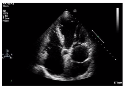
The echocardiographic image shows a mobile, highly echogenic, mesh-like structure within the right atrium consistent with the Chiari network. The Chiari network is an embryologic remnant of the right valve of the sinus venosus, appearing as a fenestrated, reticulated membrane that is usually thin and mobile, found near the orifice of the inferior vena cava or the coronary sinus.
This structure is benign and often an incidental finding but can be confused with thrombus or atrial tumors. Unlike left atrial thrombus, which appears as a more solid, immobile mass often located in the left atrial appendage, the Chiari network is mobile and located in the right atrium. Cor triatriatum is a rare congenital membrane dividing the left atrium into two chambers and appears differently on echocardiography. Artifact refers to non-anatomic echoes which do not persist or move consistently.
Recognition of Chiari network is important to avoid misdiagnosis, and its characteristics are well described in echocardiography literature such as the 'Textbook of Clinical Echocardiography' and ASE imaging guidelines16:Textbook of Clinical Echocardiography, 6ep.400-40212:ASE Guidelines on Cardiac Massesp.150-155.
Which finding is associated with partial anomalous venous return?
Partial anomalous pulmonary venous return (PAPVR) is a congenital defect where some pulmonary veins drain into the right atrium or systemic venous circulation rather than the left atrium. It is frequently associated with sinus venosus atrial septal defect (ASD), a defect near the junction of the superior vena cava and right atrium.
Cleft mitral valve is commonly associated with atrioventricular septal defects. Persistent left superior vena cava is a separate venous anomaly not typically linked with PAPVR. Perimembranous ventricular septal defects are different congenital defects not related to pulmonary venous anomalies.
The association between PAPVR and sinus venosus ASD is well described in the 'Textbook of Clinical Echocardiography, 6e', Chapter on Congenital Heart Disease and Shunt Lesions20:120-130Textbook of Clinical Echocardiography
How are pseudoaneurysms best distinguished from true aneurysms?
True ventricular aneurysms are lined by scarred myocardium and have a broad neck. Pseudoaneurysms occur after myocardial rupture contained by pericardium or scar tissue and lack myocardium in the wall. Pseudoaneurysms typically have a narrow neck and are more prone to rupture.
Pseudoaneurysms can occur at various locations, not exclusively the apex. Both true aneurysms and pseudoaneurysms may contain thrombus, so this is not a distinguishing feature.
This differentiation is important clinically and is detailed in adult echocardiography and surgical cardiology texts and ASE guidelines16:Textbook of Clinical Echocardiography, 6ep.400-40512:ASE Cardiac Masses and Aneurysms Guidelinesp.150-160.
Which two-dimensional method is recommended for assessing left ventricular ejection fraction when regional wall motion abnormalities are present?
The Simpson biplane method (method of disks) is the recommended two-dimensional echocardiographic technique to quantify left ventricular ejection fraction (LVEF), especially when regional wall motion abnormalities are present. It involves tracing endocardial borders in apical two- and four-chamber views to calculate LV volumes and EF, accounting for segmental dysfunction.
Visual estimation is subjective and less accurate. The Quinones method (single plane area-length) and Teichholz method rely on geometric assumptions and are less accurate in abnormal ventricles.
ASE chamber quantification guidelines strongly endorse Simpson biplane for LVEF assessment in regional wall motion abnormalities12:ASE Chamber Quantification Guidelinesp.70-7516:Textbook of Clinical Echocardiography, 6ep.60-65.
Get access to all 139 verified questions with detailed answers.
Unlock All AE-Adult-Echocardiography Questions